Choosing the right Airway Infarction Model begins with defining the biological question. Airway infarction is uncommon, complex, and poorly standardized in published research. A useful model must represent reduced perfusion, epithelial injury, inflammation, and repair. It should also show what happens at the tissue boundary. Otherwise, the model may look convincing but answer the wrong question.
Global respiratory disease remains a major health burden. The World Health Organization reports that chronic respiratory diseases affected hundreds of millions of people worldwide, while air pollution continues to increase respiratory risk (WHO, World Health Statistics 2024; WHO, Air Pollution and Health Reports). These figures do not directly measure airway infarction. They do show why translational airway research needs stronger disease models. Market intelligence reports also project continued growth in 3D cell culture and organ-on-chip technologies, driven by demand for more human-relevant testing (Grand View Research, 3D Cell Culture Market Report, 2024). The numbers are useful, but they cannot replace biological validation.
Donald E. Ingber, founder of the Wyss Institute, has described organs-on-chips as “microfluidic devices that recreate the physiology of living human organs.” His statement offers a practical direction for Airway Infarction Model design. A strong platform should combine human airway cells, controlled oxygen or blood-flow changes, measurable barrier damage, and repeatable imaging. Small details matter. Watch the cilia. Track lactate. Record epithelial gaps. Yet no single system is perfect. Animal models add vascular context, while organoids improve human relevance but often lack circulation. The best choice depends on the endpoint, available expertise, and evidence required. Credibility comes from comparison, not confidence.
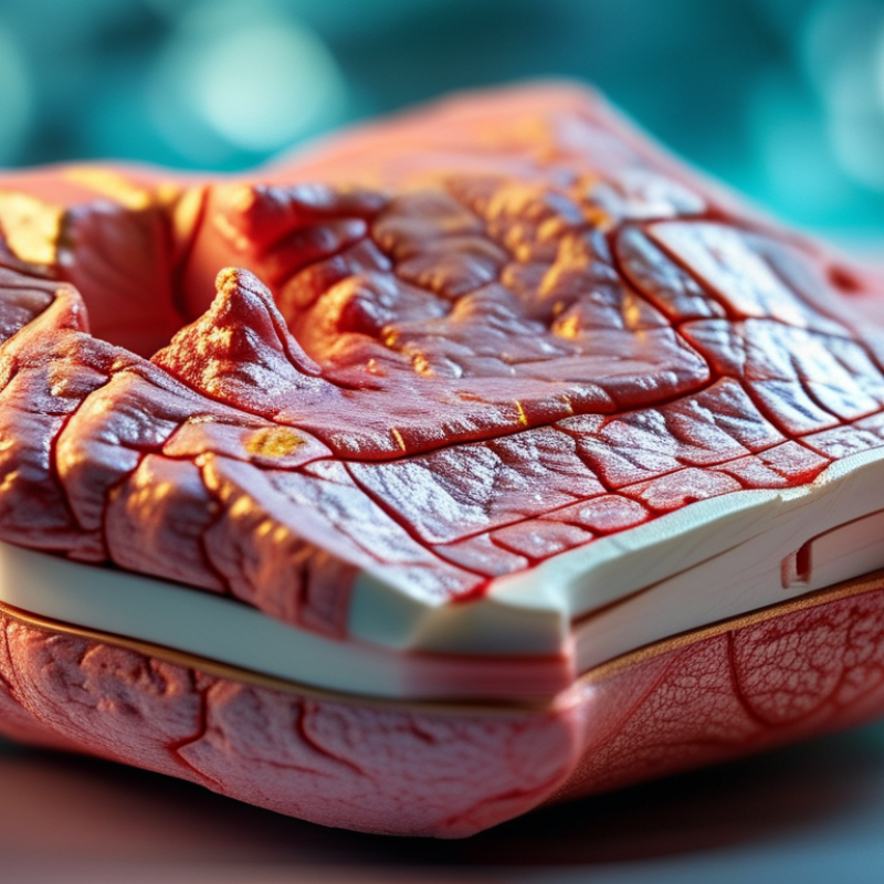
Understanding Airway Infarction and Its Research Applications
How to Choose the Right Airway Infarction Model?
Airway infarction describes tissue injury caused by inadequate blood flow to the trachea or bronchi. It is not the same as pulmonary infarction. Reduced perfusion can damage the mucosa, cartilage, and healing response. Clinically, this process may involve edema, necrosis, sloughing, stenosis, or anastomotic failure. These changes develop over time. Understanding this progression helps researchers select meaningful endpoints, rather than measuring injury through one early image.
A suitable model should match the research question. Ex vivo airway tissue can reveal epithelial damage and local vascular effects under controlled conditions. Organoid or cell-based systems may clarify barrier disruption, inflammation, and repair signals. Animal models can provide information about airway structure, blood flow, and long-term narrowing, but anatomical differences may limit translation. No model is perfect. A model may reproduce ischemia while missing immune responses or mechanical breathing forces.
Reliable study design requires defined perfusion parameters, blinded tissue assessment, and repeated observations. Useful endpoints include oxygenation, blood-flow mapping, epithelial integrity, cartilage viability, inflammatory markers, and later airway diameter. Researchers should also report anesthesia, sampling time, exclusion criteria, and welfare safeguards clearly. Small technical differences can change the apparent severity of infarction. That is easy to overlook. Combining complementary models may produce stronger evidence than relying on one elegant but incomplete system.
Defining the Scientific Goals of an Airway Infarction Model
How to Choose the Right Airway Infarction Model?
Defining the Scientific Goals of an Airway Infarction Model
An airway infarction model should begin with a precise scientific question. Are you studying reduced blood flow, epithelial death, inflammation, or tissue repair? Each goal requires different controls, exposure times, and measurable endpoints. A model built for mechanism discovery may not predict treatment response.
The clinical burden justifies careful design. The World Health Organization’s Global Health Estimates 2021 reported approximately 3.5 million COPD deaths worldwide. This figure does not represent airway infarction directly. However, it shows why airway injury research needs clinically relevant endpoints. Oxygen tension, perfusion, epithelial integrity, ciliary movement, and inflammatory markers can connect laboratory findings with patient biology.
Define the endpoint first.
An ex vivo airway model may preserve local structure and enable direct imaging. A three-dimensional culture may better represent epithelial organization. Animal models can reveal systemic responses, but species differences remain a serious limitation. That part is often underestimated. Airway diameter, immune composition, and vascular responses may not match human tissue.
Professional respiratory research standards emphasize reproducibility, transparent reporting, and biologically justified controls. Therefore, include sham injury, untreated tissue, and time-matched controls. Predefine the primary endpoint before testing multiple markers. Otherwise, attractive results may reflect selection rather than biology. A useful model is not the most complex one. It is the model that answers one question clearly, while admitting what it cannot reproduce.
Comparing Available Airway Infarction Model Types
How to Choose the Right Airway Infarction Model?
Comparing available airway infarction model types begins with the biological question. Airway infarction usually involves reduced blood flow, tissue injury, inflammation, and impaired ventilation. An ex vivo tissue model preserves airway structure and allows controlled oxygen or perfusion changes. It is useful for studying epithelial damage, but tissue viability declines quickly. An animal model adds circulation, immune responses, and breathing mechanics. However, species differences can distort drug responses and repair patterns. Organ-on-chip systems offer precise control over flow, oxygen, and mechanical stress. They are promising, though their simplified architecture may miss whole-body effects.
Disease relevance should guide model selection. The Global Initiative for Asthma’s 2024 report estimates that asthma affects over 260 million people worldwide. The World Health Organization also reports that chronic obstructive pulmonary disease caused about 3.2 million deaths in 2019. These figures support models that measure both acute injury and long-term airway remodeling. A computational model can screen oxygen gradients and vascular changes rapidly. It cannot yet reproduce living immune behavior reliably. That limitation matters.
A practical workflow may combine models. Use tissue models for early injury mapping, animal studies for systemic validation, and organ chips for human-specific mechanisms. Include oxygen saturation, epithelial integrity, vascular leakage, and airway resistance as measurable endpoints. Many studies still overvalue short-term inflammation. That may be a weakness. Model results should be compared with clinical imaging, pathology, and pulmonary-function data whenever available.
How to Choose the Right Airway Infarction Model?
Comparing Available Airway Infarction Model Types
This qualitative comparison uses a 1–5 index, where 5 indicates stronger performance for the listed criterion.
Two-dimensional cell culture generally provides high throughput but limited tissue-level relevance. Organoids and
airway-on-chip systems better reproduce three-dimensional structure and controlled vascular or inflammatory stimuli,
while animal models provide the highest whole-organism relevance but require more time, resources, and ethical oversight.
Scores are comparative planning values rather than clinical measurements.
Selecting Model Features for Accuracy and Reproducibility
Choosing an airway infarction model starts with the biological question, not the available equipment. Define whether the study examines vascular obstruction, tissue necrosis, airway narrowing, or recovery. Each endpoint requires different model features. A model with realistic airway diameter and branching can improve anatomical relevance. However, anatomical similarity alone is insufficient.
Perfusion is central to accuracy. Select a system that reproduces blood-flow changes and allows direct measurement of oxygen delivery. Tissue thickness, elasticity, and epithelial structure should also resemble the intended clinical setting. Experienced researchers should record baseline resistance, pressure, and flow before inducing injury. Small measurement errors can alter the apparent infarction size. That weakness matters.
Reproducibility depends on disciplined procedures. Use fixed injury durations, consistent anatomical locations, and predefined assessment times. Include sham or uninjured controls when ethically and scientifically appropriate. Blinded image analysis can reduce observer bias. Histology, perfusion imaging, and functional airway measurements should support one another. A single endpoint rarely tells the whole story.
Pilot testing often reveals hidden variability. Different operators may create different injury patterns, even with the same protocol. This should be reported, not quietly removed. Document animal characteristics, environmental conditions, exclusion rules, and equipment settings in detail. A reliable model is not necessarily the most complex one. It is the model that another qualified laboratory can repeat and understand. When clinical translation matters, compare model outcomes with published pathological and imaging findings rather than relying on visual similarity alone.
Validating and Refining the Chosen Airway Infarction Model
Choosing an airway infarction model is only the beginning. Validation must show that the model reflects the intended vascular injury, not merely tissue damage. Define the target airway segment, expected perfusion loss, and recovery window before testing. Use baseline imaging, oxygenation data, and tissue measurements. A visible lesion alone is not enough.
In practice, compare infarcted tissue with sham and healthy controls. Record airway pressure, blood flow, inflammatory markers, and histology at fixed time points. Look for matching evidence across methods. For example, reduced perfusion should align with epithelial injury and localized necrosis. Our first protocol looked convincing, but inconsistent sampling weakened the findings. That mistake taught us to standardize tissue depth, timing, and observer training. Inter-rater checks also matter. A clean result can still be misleading.
Tips: Pilot the model with a small, ethically approved study. Confirm that the injury is localized and reproducible. Test whether anesthesia, ventilation, or handling changes the outcome. Keep raw images and exclusion decisions. Do not remove unusual results without a documented reason. If the model fails to reproduce expected airway changes, refine the induction method or select a better endpoint. Reliability grows through transparent adjustments, not perfect-looking data.
FAQS
: Define one main question. Study perfusion loss, epithelial death, inflammation, narrowing, or repair. Do not combine every endpoint casually.
Ex vivo tissue supports local imaging. Three-dimensional cultures show epithelial organization. Animal models reveal systemic responses, but species differences can weaken translation.
Choose realistic airway diameter, branching, tissue thickness, elasticity, and epithelial structure. Measure blood flow and oxygen delivery directly whenever possible.
Include sham-injured, untreated, healthy, and time-matched controls when appropriate. Predefine the primary endpoint before collecting many measurements.
Combine perfusion imaging, oxygenation data, histology, inflammatory markers, and airway function. Reduced perfusion should match localized necrosis and epithelial damage.
Keep injury duration, anatomical location, and assessment times consistent. Record baseline pressure, resistance, and flow. Blinded image analysis can reduce observer bias.
Operators may create different injury patterns. Anesthesia, ventilation, handling, tissue depth, and sampling time can also change outcomes. That weakness matters.
Keep raw images and document exclusion decisions. Do not remove unusual findings without a clear reason. Our early protocol looked convincing, but sampling inconsistency weakened it.
Confirm localized, reproducible injury across independent methods. Compare results with pathological and imaging evidence. A visible lesion alone is not enough.
Run a small, ethically approved pilot study. If expected airway changes do not appear, adjust the induction method or choose a clearer endpoint. Perfect data can still mislead.
Conclusion
Choosing the right Airway Infarction Model begins with understanding the biological mechanisms, clinical characteristics, and research applications of airway tissue injury caused by impaired blood flow. Researchers should first define the main scientific objectives, such as investigating disease progression, evaluating therapeutic strategies, studying tissue repair, or identifying relevant biomarkers. These goals will determine whether an in vitro, ex vivo, or in vivo model is most appropriate and which physiological features must be represented.
A reliable model should balance biological accuracy, practicality, ethical considerations, and reproducibility. Important selection factors include the similarity of airway structure, control of injury severity, consistency of experimental conditions, measurable outcomes, and compatibility with available analytical methods. After selecting a model, researchers should validate it against established pathological, molecular, and functional indicators. Continuous refinement based on experimental results can improve reliability and ensure that the Airway Infarction Model remains suitable for the intended research purpose.

 Guanbang Medical